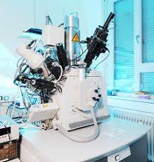
Dual Beam FIB
A Focused Ion Beam (FIB) instrument uses a finely focused ion beam to modify and image the sample of interest. FIB is chiefly used to create very precise cross sections of a sample for subsequent imaging via SEM, STEM or TEM or to perform circuit modification. Additionally, FIB imaging can be used to image a sample directly, detecting emitted electrons either from the ion or electron beam. The contrast mechanism for FIB is different than for SEM or S/TEM, so unique structural information can be obtained in some cases. A dual beam FIB/SEM integrates these two techniques into one tool whereas a single beam FIB contains only an ion beam, with electron beam imaging taking place in a separate SEM, STEM or TEM instrument.
As a sample preparation tool, the FIB can accurately produce cross-sections of a sample that are impossible to create otherwise:
FIB analysis has revolutionized sample preparation for TEM samples, making it possible to identify sub-micron features and precisely prepare cross sections.
- FIB-prepared sections are used extensively in SEM microscopy, where the FIB preparation, SEM imaging, and elemental analysis can happen with the same multi-technique tool.
- FIB-prepared sections are also used in Auger Electron Spectroscopy to provide elemental identification of subsurface features quickly and precisely.
- It is an ideal tool for examining products with small, difficult-to-access features, such as those found in the semiconductor industry and for sub-surface particle identification.
- It is a good option for products that are hard to cross section, such as a soft polymer that is challenging to polish.
Ideal Uses of FIB
- SEM, STEM and TEM sample preparation
- High resolution cross-section images of small, hard-to-access sample features
- Micro sampling via in-situ liftout
Strengths
- Best method to cross-section small targets
- Rapid, high-resolution imaging
- Good grain contrast imaging
- Versatile platform that supports many other tools
Limitations
- Vacuum compatibility typically required
- Imaging may spoil subsequent analyses
- Residual Ga on analytical face
- Ion beam damage may limit image resolution
- Cross-section area is small
FIB Technical Specifications
- Signals Detected: Electrons, secondary ions, X-rays, light (Cathodoluminescence)
- Imaging/Mapping: Yes
- Lateral Resolution/Probe Size: 7 nm (ion beam); 20 nm (electron beam)